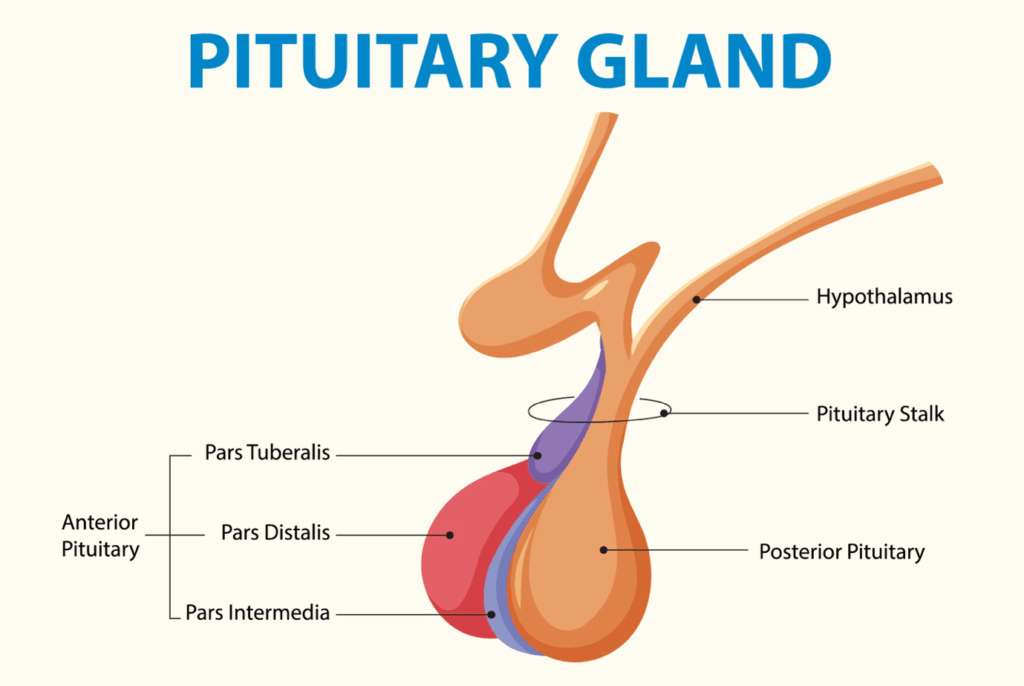
A graphic showing the parts of the pituitary gland.

Pituitary Tumors: When Is Surgery the Answer?

A graphic showing the parts of the pituitary gland.

The pituitary gland, a diminutive but indispensable endocrine gland nestled at the base of the brain, plays a pivotal role in regulating a myriad of essential bodily functions. This master gland secretes a variety of hormones that govern growth, metabolism, reproduction, stress response, and fluid balance.

Pituitary tumors, abnormal growths that develop on the pituitary gland, can disrupt the delicate hormonal balance orchestrated by this gland, leading to a spectrum of health complications. While not all pituitary tumors necessitate surgical intervention, surgery remains a critical treatment modality in specific clinical scenarios.

Understanding Pituitary Tumors

Pituitary tumors are predominantly benign, i.e., they are noncancerous growths that do not typically spread to other parts of the body. However, their location and their potential to interfere with pituitary function make them a significant health concern.

Pituitary tumors are classified into two main categories, based on their hormonal activity:

  • Functioning Tumors:These tumors are characterized by their ability to secrete excess amounts of one or more pituitary hormones.
  • Nonfunctioning Tumors:These tumors, in contrast, do not secrete excess hormones. They may remain asymptomatic for extended periods and are often discovered incidentally during imaging studies performed for other reasons.

The Diagnostic Pathway for Pituitary Tumors

The diagnosis of a pituitary tumor typically involves a combination of clinical evaluation, hormonal testing, and advanced imaging techniques:

  • Hormone Tests:Blood and urine tests are essential to measure the levels of various pituitary hormones and identify any hormonal excesses or deficiencies.
  • Imaging Tests:Magnetic resonance imaging (MRI) is the gold standard imaging modality for visualizing the pituitary gland and detecting pituitary tumors.
  • Vision Tests:Visual field testing is performed to assess for any visual impairments caused by compression of the optic nerves.

When Is Surgery the Answer?

A woman in a white dress holds her hands to her head as though she has a headache.
One of the symptoms of pituitary tumors is headache.

The decision to pursue surgery for a pituitary tumor is a complex one, requiring careful consideration of several factors.

Surgery is typically considered when:

  • The Tumor Is Causing Significant Symptoms:When the tumor is responsible for bothersome or debilitating symptoms, such as persistent headaches, vision problems, or hormonal imbalances that are not adequately controlled with medication, surgery may be recommended to alleviate these symptoms.
  • The Tumor Is Compressing the Optic Nerves and Threatening Vision:Compression of the optic nerves poses a serious risk to vision. Surgery is often necessary to decompress the optic nerves and prevent or reverse vision loss.
  • The Tumor Is a Functioning Tumor That Is Producing Excess Hormones:In cases where a functioning tumor is secreting excess hormones and causing significant health problems, surgery may be performed to remove the tumor and restore normal hormone levels.

Surgical Approaches to Pituitary Tumors

The surgical approach to a pituitary tumor depends on its size, location, and relationship to surrounding structures.

The two main surgical techniques are:

  • Transsphenoidal Surgery:This is the most common and preferred surgical approach for most pituitary tumors. It is a minimally invasive procedure that involves accessing the pituitary gland through the nasal passages and sphenoid sinus (an air-filled space behind the nasal cavity).
  • Craniotomy:In rare cases, a craniotomy, which involves opening the skull, may be necessary for very large or complex tumors that cannot be safely removed through the transsphenoidal approach.

Seeking Expert Care

If you have been diagnosed with a pituitary tumor or are experiencing symptoms that may suggest a pituitary tumor, it is important to seek evaluation and treatment from a specialized medical center with expertise in pituitary disorders.

The physicians at Houston Physicians Hospital can help.

On the page of Neurosurgery and Spine Specialist in Webster, you can scroll down and find a physician who’s right for you. Then click their link to visit their website and make an appointment.

Biofeedback therapy uses sensors to monitor pelvic floor muscle activity, helping patients gain control over these muscles.

The Science of Self-Regulation: How Biofeedback Is Used in Pelvic Floor Therapy

 Biofeedback therapy uses sensors to monitor pelvic floor muscle activity, helping patients gain control over these muscles.

The pelvic floor, a complex network of muscles, ligaments, and connective tissue forms a supportive sling across the base of the pelvis. This structure plays a vital role in supporting the pelvic organs, including the bladder, bowel, uterus (in women), and prostate gland (in men).

Beyond organ support, the pelvic floor is essential for maintaining continence, facilitating sexual function, and contributing to core stability. Dysfunction in this intricate system can lead to a spectrum of challenging conditions, significantly impacting an individual’s quality of life.

Pelvic floor therapy offers a range of interventions to address these issues, and biofeedback has emerged as a sophisticated and effective tool within this specialized field.

Biofeedback: A Technology-Driven Approach to Retraining

Biofeedback is a noninvasive therapeutic technique that empowers individuals to develop heightened awareness and voluntary control over their physiological processes.

In the context of pelvic floor therapy, biofeedback employs specialized sensors to monitor the activity of the pelvic floor muscles. These sensors, which may be surface electrodes or internal sensors, detect muscle contractions and relaxation. The information gathered is then translated into a user-friendly format.

This immediate and objective feedback loop is instrumental in helping patients learn to identify, isolate, contract, and relax their pelvic floor muscles correctly.

The Science of Self-Regulation

Biofeedback operates on the principles of operant conditioning, a fundamental learning theory that posits that behavior is modified by its consequences. By providing immediate and contingent feedback on muscle activity, biofeedback reinforces desired muscle responses.

For instance, if a patient is attempting to contract their pelvic floor muscles, the biofeedback display will provide positive reinforcement when the contraction is performed correctly, encouraging repetition of that behavior. Conversely, if the patient is contracting the wrong muscles or not contracting effectively, the feedback will indicate this, allowing them to adjust their efforts.

Through consistent practice and repetition, patients can gradually refine their muscle control and achieve greater self-regulation of their pelvic floor function.

Applications of Biofeedback in Pelvic Floor Therapy

 A woman doing exercises with her baby.
Biofeedback therapy uses sensors to monitor pelvic floor muscle activity, helping patients gain control over these muscles.

Biofeedback has demonstrated efficacy in addressing a wide spectrum of pelvic floor conditions:

  • Incontinence:Biofeedback is a valuable tool for individuals experiencing urinary or fecal incontinence. By providing visual or auditory feedback on muscle activity, it helps patients learn to identify and strengthen the specific muscles that control the flow of urine or stool.
  • Pelvic Pain:For individuals grappling with chronic pelvic pain, often associated with hypertonic or overly tight pelvic floor muscles, biofeedback can facilitate muscle relaxation techniques. The feedback allows patients to visualize their muscle tension and learn to consciously release it.
  • Constipation:Biofeedback can be employed to improve the coordination of muscles involved in bowel movements. By providing feedback on muscle activity during defecation, it can help patients learn to relax the appropriate muscles and facilitate effective bowel evacuation.
  • Sexual Dysfunction:Biofeedback can be a component of treatment for certain types of sexual dysfunction, such as dyspareunia (painful intercourse) or vaginismus (involuntary muscle spasms of the vagina). By enhancing muscle function and awareness, it can contribute to improved sexual comfort and function.

Recommended Read: Understanding Pelvic Floor Disorders in Women

The Multifaceted Benefits of Biofeedback in Pelvic Floor Therapy

Biofeedback offers a range of advantages in the treatment of pelvic floor dysfunction:

  • Enhanced Awareness:Biofeedback provides patients with a unique and objective window into their pelvic floor muscle activity, fostering a deeper understanding of how these muscles function and how they contribute to various bodily processes.
  • Improved Muscle Control:Through the real-time feedback provided by biofeedback, patients can develop more precise and effective control over their pelvic floor muscles, learning to contract and relax them with greater accuracy and coordination.
  • Personalized Treatment:Biofeedback protocols can be tailored to the specific needs and challenges of each individual, allowing for a personalized and targeted approach to therapy.

Taking the First Step

If you are experiencing symptoms of pelvic floor dysfunction, such as incontinence, pelvic pain, or sexual problems, pelvic floor therapy with biofeedback may offer a viable and effective treatment option.

The physicians at Houston Physicians Hospital specialize in pelvic health. They can provide an accurate diagnosis and discuss appropriate treatment strategies.

On the Pelvic Floor Therapy page, you can scroll down and find a physical therapy location near you. Then click on the location to make an appointment.

A man smiling in a hospital bed, recovering after a successful deep brain stimulation (DBS) procedure.

DBS for Parkinson’s: A Promising Treatment Option

A man smiling in a hospital bed, recovering after a successful deep brain stimulation (DBS) procedure.

Parkinson’s disease, a progressive neurological disorder, affects movement, causing tremors, stiffness, slowness, and balance problems. While medication can manage symptoms in the early stages, their effectiveness can decrease over time, and side effects can become challenging.

Deep brain stimulation (DBS) has emerged as a promising treatment option for individuals with Parkinson’s disease who are experiencing motor fluctuations and other complications.

Understanding Parkinson’s Disease: A Disruption in Movement

Parkinson’s disease is characterized by the loss of dopamine-producing cells in a specific area of the brain called the substantia nigra. Dopamine is a neurotransmitter that plays a crucial role in controlling movement. As dopamine levels decline, the hallmark symptoms of Parkinson’s disease appear.

Deep Brain Stimulation: A Neurological Intervention

DBS is a surgical procedure that involves implanting a small, pacemaker-like device that sends electrical impulses to specific areas of the brain that control movement. These electrical impulses help regulate the abnormal brain activity that causes Parkinson’s disease symptoms.

DBS does not cure Parkinson’s disease, but it can significantly reduce symptoms and improve quality of life for carefully selected individuals.

The procedure involves the following steps:

  1. Brain Mapping:Before surgery, detailed brain imaging, such as MRI, is used to identify the precise brain targets for stimulation. These targets are typically the subthalamic nucleus (STN) or the globus pallidus interna (GPi), both of which are involved in movement control.
  2. Electrode Implantation:During surgery, thin wires called electrodes are implanted into the targeted brain regions. The procedure is typically performed while the patient is awake to allow for neurological testing to ensure accurate placement.
  3. Pulse Generator Implantation:A small pulse generator, similar to a pacemaker, is implanted under the skin in the chest. This generator sends electrical impulses to the electrodes in the brain.
  4. Connecting Wires:Wires are tunneled under the skin to connect the electrodes in the brain to the pulse generator in the chest.
  5. Programming:After surgery, the pulse generator is programmed to deliver the appropriate electrical stimulation. The settings are adjusted over time to optimize symptom control and minimize side effects.

Benefits of DBS for Parkinson’s Disease

DBS can provide significant benefits for individuals with Parkinson’s disease, including:

  • Reduced Tremors:DBS is highly effective in reducing tremors, which are often a debilitating symptom of Parkinson’s disease.
  • Decreased Rigidity and Stiffness:DBS can help alleviate muscle stiffness and rigidity, making movement easier.
  • Improved Bradykinesia (Slowness of Movement):DBS can improve bradykinesia, allowing for smoother and faster movements.
  • Reduced Dyskinesias (Involuntary Movements):DBS can help reduce dyskinesias, which are involuntary movements caused by long-term use of Parkinson’s medications.
  • Decreased Medication Needs:DBS can often allow individuals to reduce their dosage of Parkinson’s medications, which can, in turn, reduce medication-related side effects.
  • Improved Quality of Life:By reducing symptoms and improving function, DBS can significantly improve the quality of life for individuals with Parkinson’s disease.

Who Is a Candidate for DBS?

A man being prepared for deep brain stimulation (DBS) surgery.
Candidates for DBS typically have Parkinson’s disease that responds to medication but are experiencing motor fluctuations or side effects.

DBS is not suitable for everyone with Parkinson’s disease.

Ideal candidates typically:

  • Have Parkinson’s disease that is responsive to levodopa, a common Parkinson’s medication.
  • Experience motor fluctuations, e.g., “on-off” periods, when medication effectiveness wears off.
  • Develop dyskinesiasthat are difficult to manage with medication.
  • Are in relatively good overall health and can tolerate surgery.

Your Partner in Neurological Health

The physicians at Webster Surgical Hospital are committed to providing quality neurological care for individuals with Parkinson’s disease and other movement disorders. They offer advanced Parkinson’s treatment in Houston, including deep brain stimulation, to help patients manage their symptoms and improve their quality of life.

On the Deep Brain Stimulation page, you can scroll down and find a physician who’s right for you. Then click their link to visit their website and make an appointment.

A woman holding her head and showing signs of distress, indicating psychogenic pain.

Psychogenic vs. Physical Pain: Understanding the Difference

A woman holding her head and showing signs of distress, indicating psychogenic pain.

We typically associate pain with a clear physical cause: a cut, a bruise, a broken bone. But pain isn’t always that straightforward. Sometimes, pain can arise or persist without an identifiable physical injury or disease. This is where the concept of psychogenic pain comes into play.

Understanding the distinction between psychogenic pain and physical pain is crucial for effective diagnosis and treatment. Both types of pain are real and can be debilitating, but their origins and management strategies differ significantly.

Let’s take a closer look.

The Nature of Physical Pain: A Clear Connection

Physical pain, also known as nociceptive pain, is the type of pain we typically think of. It arises from tissue damage or inflammation.

When you stub your toe, for example, specialized sensory receptors called nociceptors send signals through the nervous system to the brain, which interprets these signals as pain.

Key characteristics of physical pain include:

  • Identifiable Cause:Physical pain usually has a clear and identifiable cause, e.g., an injury, infection, or disease.
  • Location:The pain is typically localized to the site of the injury or inflammation.
  • Consistency:The pain pattern is often consistent with the nature of the injury or condition. For example, pain from a broken bone worsens with movement.
  • Response to Treatment:Physical pain often responds well to treatments that address the underlying cause, e.g., pain relievers, anti-inflammatory medications, or surgery.

The Enigma of Psychogenic Pain: Mind and Body Intertwined

Psychogenic pain, also known as psychogenic disorder pain or functional pain, is a more complex phenomenon. It’s characterized by pain that is significantly influenced by psychological factors, such as thoughts, emotions, and behaviors.

While the pain is real to the person experiencing it, there may not be a clear physical cause or the pain may be disproportionate to any identifiable physical findings.

It’s important to emphasize that psychogenic pain is not “all in the head” or imagined. The pain is a genuine experience, but its origins and modulation are significantly affected by psychological processes.

Key characteristics of psychogenic pain include:

  • Lack of Clear Physical Cause:There may not be a readily identifiable physical injury, disease, or inflammation that adequately explains the intensity or persistence of the pain.
  • Disproportionate Pain:The pain may be disproportionate to any physical findings. For example, a minor injury may trigger intense or chronic pain.
  • Fluctuating or Variable Pain:The pain may fluctuate in intensity or location, and it may be influenced by stress, emotional state, or psychological factors.
  • Psychological Factors:Psychological factors, such as anxiety, depression, stress, trauma, and unresolved emotional issues play a significant role in the experience of pain.
  • Limited Response to Traditional Pain Treatments:Psychogenic pain may not respond well to traditional pain treatments that target physical causes.

Identifying Psychogenic Pain

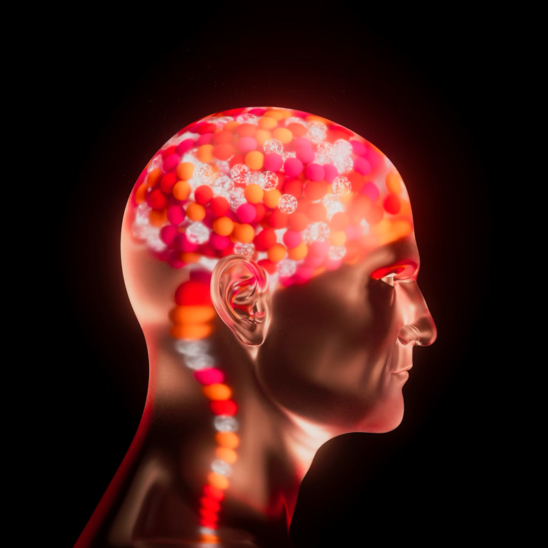
An illustration depicting heightened brain activity, emphasizing the role of the central nervous system in processing psychogenic pain.
https://unsplash.com/photos/a-womans-head-with-colorful-beads-on-it–AazHiQpS-A

Diagnosing psychogenic pain requires a careful and comprehensive evaluation. There is no single test to definitively diagnose psychogenic pain.

The evaluation typically involves:

  • Medical History and Physical Examination:The physician will gather a detailed medical history and perform a thorough physical examination to rule out any underlying physical causes of the pain.
  • Psychological Assessment:A psychological assessment may be conducted to evaluate the patient’s mental and emotional state, including anxiety, depression, stress, and trauma history.
  • Pain Assessment:A detailed pain assessment will be conducted to characterize the pain, including its location, intensity, quality, and pattern.
  • Functional Assessment:An assessment of how pain affects the patient’s daily functioning and activities.

Addressing the Complexities of Pain

Understanding the difference between psychogenic and physical pain is essential for effective diagnosis and treatment. Psychogenic pain is a real and often debilitating condition that requires a multidisciplinary approach that addresses the interplay between the mind and body. By recognizing the role of psychological factors in the pain experience, healthcare providers can provide more comprehensive and effective care, helping individuals find relief and improve their quality of life.

Your Partner in Pain Management

The physicians at Houston Physicians Hospital understand the complexities of pain and the importance of addressing both the physical and psychological aspects of pain management. They offer a range of treatment options, including psychotherapy referrals and physical therapy services, to help patients find relief and improve their overall well-being.

On the Pain Management page, you can scroll down and find a physician who’s right for you. Then click their link to visit their website and make an appointment.

A man experiencing discomfort related to neurogenic bladder.

Common Causes of Neurogenic Bladder

A man experiencing discomfort related to neurogenic bladder.

The bladder, a seemingly simple organ, plays a vital role in our daily lives. Its ability to store and eliminate urine efficiently is essential for comfort, health, and social well-being. This process is orchestrated by a complex communication network between the brain, the spinal cord, and the bladder itself. When this communication network is disrupted due to neurological conditions, it can lead to a condition known as neurogenic bladder.

Neurogenic bladder is not a single disease entity, but rather a term that encompasses a range of bladder dysfunctions caused by damage to the nerves that control the bladder. This disruption can manifest in various ways, affecting the bladder’s ability to store, hold, and eliminate urine. Understanding the causes, impact, and management of neurogenic bladder is imperative.

Common Causes of Neurogenic Bladder

A variety of neurological conditions can cause or contribute to neurogenic bladder.

The more common causes include:

  • Spinal Cord Injury:Damage to the spinal cord is a frequent cause of neurogenic bladder. The location and severity of the injury determine the type and extent of bladder dysfunction. Injuries in the upper spinal cord typically lead to overactive bladder, while injuries in the lower spinal cord may result in an underactive bladder.
  • Multiple Sclerosis (MS):This autoimmune disease affects the central nervous system and can damage the myelin sheath, the protective covering of nerve fibers. This damage can disrupt the nerve signals that control bladder function.
  • Stroke:A stroke, caused by a disruption of blood flow to the brain, can damage brain areas that control bladder function. This damage can result in various bladder problems, including incontinence, urgency, and frequency.
  • Parkinson’s Disease:This progressive neurological disorder affects movement and can also impact bladder control. Symptoms may include urinary urgency, frequency, and incontinence.
  • Diabetes:Over time, diabetes can damage nerves throughout the body, including those that control the bladder. This diabetic neuropathy can lead to bladder dysfunction.
  • Brain Tumors or Injury:Tumors or traumatic injuries to the brain can disrupt the nerve pathways that control bladder function. The specific bladder problems depend on the location and extent of the damage.
  • Congenital Conditions:Some individuals are born with neurological conditions that can cause neurogenic bladder. Spina bifida, a birth defect that affects the spinal cord, is a common example.

Diagnosis and Management

Diagnosing neurogenic bladder involves a comprehensive evaluation to identify the underlying cause and assess the extent of bladder dysfunction.

This evaluation may include:

  • Medical History Review:The physician will review the patient’s medical history, including any neurological conditions or injuries.
  • Physical Examination:A physical exam may be performed to assess neurological function and bladder function.
  • Neurological Examination:This may include tests to assess reflexes, sensation, and muscle strength.
  • Urodynamic Testing:These tests measure bladder function, including bladder capacity, bladder pressure, and urine flow rate.
  • Imaging Studies:Imaging tests, such as ultrasound or cystoscopy, may be used to visualize the bladder and urinary tract.

The management of neurogenic bladder is tailored to the individual patient’s needs and the underlying cause of the condition.

Treatment options may include:

  • Medication:Medication can help manage symptoms like overactive bladder or urinary retention.
  • Catheterization:Intermittent catheterization, where a thin tube is inserted into the bladder to drain urine, may be necessary for individuals who are unable to empty their bladder effectively.
  • Bladder Training:This involves techniques to improve bladder control, such as timed voiding and pelvic floor exercises.
  • Pelvic Floor Exercises:These exercises strengthen the muscles that support the bladder and can improve bladder control. Pelvic floor therapy may also be recommended.
  • Surgery:In some cases, surgery may be necessary to improve bladder function or address complications.

Houston Physicians’ Hospital: A Focus on Improving Lives

A man smiling in a hospital bed after receiving treatment for neurogenic bladder.
Houston Physicians’ Hospital offers quality care and a range of treatment options for individuals with neurogenic bladder.

The physicians at Houston Physicians Hospital understand the challenges that neurogenic bladder can present. Their doctors are dedicated to providing quality care and support to individuals with this condition.

They offer a range of diagnostic and treatment options to help patients manage their symptoms, improve their bladder function, and enhance their quality of life.

On the Urology Care page, you can scroll down and find a physician who’s right for you. Then click their link to visit their website and make an appointment.

A person exhibiting signs of a stroke.

Recognizing the Symptoms of a Stroke and What to Do Next

A person exhibiting signs of a stroke.

A stroke, sometimes called a “brain attack,” is a serious medical emergency that occurs when blood flow to the brain is interrupted. This interruption can be caused by a blocked artery (ischemic stroke) or a ruptured blood vessel (hemorrhagic stroke). When a stroke occurs, brain cells begin to die due to lack of oxygen, which can lead to permanent brain damage, disability, or even death.

Recognizing the symptoms of a stroke and acting quickly are crucial for minimizing brain damage and improving the chances of a full recovery.

Understanding the Urgency

During a stroke, every minute counts. The longer the brain is deprived of oxygen, the greater the potential for damage. Prompt medical intervention can help restore blood flow to the brain and limit the extent of the damage.

The FAST Acronym: A Lifesaving Tool

The American Stroke Association has developed an easy-to-remember acronym called FAST to help people recognize the most common symptoms of a stroke:

  • Face Drooping: Does one side of the face droop, or is it numb? Ask the person to smile.
  • Arm Weakness: Is one arm weak or numb? Ask the person to raise both arms. Does one arm drift downward?
  • Speech Difficulty: Is speech slurred? Is the person unable to speak or hard to understand? Ask the person to repeat a simple sentence, like “The sky is blue.”
  • Time to Call 911: If someone shows any of these symptoms, even if the symptoms go away, call 911 immediately.

Other Potential Stroke Symptoms

In addition to the FAST signs, other symptoms of a stroke can include:

  • Sudden numbness or weakness of the leg. This can manifest as a loss of feeling or an inability to move the leg.
  • Sudden confusion; trouble understanding. This can involve difficulty processing information, understanding spoken or written language, or following simple instructions.
  • Sudden trouble seeing in one or both eyes. This can range from blurred vision or double vision to a partial or complete loss of vision in one or both eyes.
  • Sudden trouble walking, dizziness, loss of balance or coordination. The person may feel dizzy or unsteady, or have a sense of vertigo.
  • Sudden severe headache with no known cause. This is often described as a “thunderclap” headache, which is an intense headache that comes on suddenly and reaches peak intensity within seconds or minutes.

What to Do Next: Acting Quickly Can Save Lives  

If you suspect someone is having a stroke, take the following steps:

  1. Call 911 Immediately:Do not delay. Emergency medical technicians are trained to provide immediate care and transport the person to the hospital quickly.
  2. Note the Time:Try to determine when the symptoms first started. This information is crucial for medical professionals to determine the best course of treatment.
  3. Do Not Drive the Person to the Hospital Yourself:Emergency medical technicians can provide necessary medical care en route to the hospital.
  4. Keep the Person Calm and Comfortable:Reassure the person and keep them as comfortable as possible while waiting for emergency medical services.
  5. Do Not Give the Person Anything to Eat or Drink:This could lead to choking.

Treatment Options: Restoring Blood Flow

Treatment for stroke depends on the type of stroke. Ischemic strokes are often treated with clot-busting drugs, such as tPA (tissue plasminogen activator), which can dissolve the clot and restore blood flow to the brain. Hemorrhagic strokes may require surgery to stop the bleeding.

A woman smiling in a hospital bed, recovering after receiving stroke treatment.
Prompt medical intervention and rehabilitation are essential for stroke recovery and regaining lost function.

After a stroke, rehabilitation is often necessary to help individuals regain lost function. Rehabilitation may include physical therapy, occupational therapy, and speech therapy.

Your Partner in Neurological Care

The physicians at Houston Physicians Hospital provide quality neurological care. They’re trained to diagnose and treat strokes, and they offer comprehensive rehabilitation services to help patients on their road to recovery.

On the Neuroscience Rehabilitation page, you can scroll down and find a neurosurgeons in Clear Lake TX, who’s right for you. Then click their link to visit their website and make an appointment.